
Cardiology
Cardiology
The cardiology department treats coronary diseases (24-hour coronary emergencies), rhythm and conduction disorders, heart failure and valvular, muscular and malformative diseases in adults and children from the 3 departments of Western Normandy.
The cardiology department treats coronary diseases (24-hour coronary emergencies), rhythm and conduction disorders, heart failure and valvular, muscular and malformative diseases in adults and children from the 3 departments of Western Normandy.

Consultations & Team
Service composition
- 2 University Professors · Hospital Practitioners
- 2 University Lecturers · Hospital Practitioners
- 13 Hospital Practitioners
- 4 University Clinical Heads, Hospital Assistants
- 6 Attached Practitioners
- 3 Health Executives

Pathologies

Atrial Septal Defect (ASD)
An atrial septal defect (ASD) is a congenital heart defect characterized by a defect in the formation of the interatrial septum (the wall separating the upper chambers of the heart, namely the left and right atria), which can vary in shape and size. This abnormal hole results in excessive blood flow from the left atrium (high pressure) to the right atrium (low pressure), a phenomenon known as a shunt. Because the blood pressure on the left side of the heart is higher than on the right, blood constantly flows into the right atrium through this opening, potentially causing dilation of the right atria and heart chambers. Over time, this defect increases the risk of pulmonary hypertension, heart failure, arrhythmias, and stroke. The indication for ASD closure is considered in cases of significant shunt, right ventricular dilation, and the appearance of symptoms such as dyspnea or decreased physical capacity.
Thanks to the development of percutaneous techniques, closure of atrial septal defects (ASDs) using a catheter-introduced occlusion device is now the most common technique. The operator inserts a closure device through the femoral vein in the groin. The folded device, introduced into the heart via the catheter, expands to close the ASD. Small residual shunts that persist after the procedure often close over time as endothelial tissue covers the device. Increasingly, ASD closure by interventional cardiac catheterization is considered a routine treatment requiring a very short hospital stay and with a straightforward recovery.
The Patent Foramen Ovale (PFO)
A patent foramen ovale (PFO) is defined as the persistence, after birth, of a communication between the two atria, a kind of tunnel potentially responsible for the passage of venous blood into arterial blood within the heart. Normally, this septum is closed at birth but can sometimes present a small opening called the foramen ovale. This communication, essential during fetal life, closes after birth in the vast majority of cases.
Unlike ASDs, this is not an opening due to a lack of substance, but rather a failure of adhesion of the primum and secondum, which form the interatrial septum of the adult heart.
When the PFO remains patent with a right-to-left shunt, associated with an aneurysm of the interatrial septum, it may be a contributing factor to stroke (CVA).
The examination that allows identification of the PFO is transesophageal echocardiography (TEE) with a bubble test showing the passage of microbubbles into the left atrium after injection via a peripheral venous route.
Transesophageal echocardiography (TEE) allows for the evaluation of the anatomy of the interatrial septum and the identification of an aneurysm. A patent foramen ovale (PFO) can cause arterial embolism, unexplained and recurrent strokes, hypoxia uncorrectable with oxygen, decompression syndrome in divers, and migraines unrelieved by treatment. Its discovery may lead to discussion of percutaneous closure using a prosthesis comprising two discs (several prosthesis models are available) ensuring complete closure of the shunt.
THE RISKS?
As with any cardiac and vascular procedure, complications are possible. These complications are most often minor. The expected long-term benefits far outweigh the risks associated with these procedures.
Transcatheter aortic valve implantation (TAVI)
Aortic stenosis is a disease that affects older people, particularly men over 70. The opening of the aorta narrows, hindering the ejection of blood from the left ventricle. When the obstruction is critical, intervention is necessary if it is causing symptoms you are experiencing (shortness of breath, chest tightness, dizziness, fatigue, etc.) because there is a risk of hospitalization and death.
Atrial (or atrial) fibrillation ablation
This is the most complex arrhythmia. It is not organized and regular, but rather the result of very rapid, disordered, and chaotic electrical activation of the atria. Unlike most other tachycardias, its ablation cannot be reduced to the destruction of a single target (such as an electrical source or an area of abnormal conduction). It is an electrical abnormality affecting the entire atrial tissue that must be treated. The areas most frequently involved are usually located in the left atrium (LA), and more specifically at the junction of the LA and the pulmonary veins. Over time and with repeated episodes of fibrillation, certain areas of the atria become damaged and themselves become sources of fibrillation. In some cases, these areas require treatment in addition to that of the pulmonary veins.
Atrial fibrillation progresses over time, typically leading to increasingly prolonged episodes. It can cause serious complications, which vary depending on the individual patient's underlying health and risk factors. The most significant complication is the risk of stroke due to impaired drainage of the atrial chambers during fibrillation, resulting in blood pooling and the potential for clotting. Fibrillation can also lead to all stages of heart failure, from simple fatigue or shortness of breath on exertion to severe damage to the heart muscle with serious signs of heart failure.
Overall, atrial fibrillation is estimated to cause mortality comparable to that of myocardial infarction, diabetes, or hypertension. Beyond the serious prognostic implications of the condition, the effects of the symptoms associated with the arrhythmia on mood and quality of life can be as significant as those of a serious illness such as cancer.
Treatment varies but essentially consists of either compartmentalizing the atria with radiofrequency lines, or selectively treating areas that appear pathological and responsible for maintaining the arrhythmia.
Radiofrequency ablation: During the procedure, performed under anesthesia, a probe is inserted into the femoral vein of the lower limb and then guided to the heart. The tip of the probe is used to ablate small areas of the heart responsible for the arrhythmia.
The cavities traversed by the catheter are reconstructed in 3D by a specific computer system, which then allows the probes to be moved and the areas to be treated to be identified with very high precision. The operation aims for a true electrical disconnection between the atrium and the ventricle.
After the procedure and waking up, post-operative monitoring in a continuous care unit may be necessary, but hospitalization is generally short, from 2 to 3 days.
Percutaneous closure of the left auricle
Atrial fibrillation (AF) is the most common cardiac arrhythmia and carries a significant cardioembolic risk that increases considerably with age. Indeed, irregular heartbeats can lead to the formation of blood clots (or thrombi). These clots can then travel through the bloodstream and block an artery, particularly in the brain: this is a stroke.It turns out that the thrombi responsible for thromboembolic events in non-valvular AF form preferentially in the left atrium in more than 90% of cases. This appendage, located on the surface of the left atrium, is shaped like a small pouch or protrusion, varies from person to person, and has no specific function.
In cases of atrial fibrillation, anticoagulant treatments aim to thin the blood to prevent clot formation and thus the risk ofstroke. However, they can cause bleeding, sometimes severe. Occlusion of the appendix, using a percutaneous closure system with a special prosthesis, offers an alternative to anticoagulant treatment for individuals at high risk of bleeding.
This procedure is performed under general anesthesia via a percutaneous approach. The device is delivered to the right atrium by catheterization, through puncture of the right femoral vein (in the groin). Then, by puncture of the transseptal wall (the septum between the two atria) under echocardiographic and/or fluoroscopic guidance, the left atrium is accessed as far as the left atrial appendage. The prosthesis is then implanted at the neck of the atrial appendage: first, the distal portion is deployed, positioning itself in the neck of the appendage like a plug; then, the gradual withdrawal of the delivery catheter releases the proximal disc of the prosthesis, which expands over the opening of the atrial appendage and covers it.
Once the prosthesis is in place, blood clots can no longer migrate from this auricle, and the risk ofstroke is significantly reduced. Anticoagulant treatment can then be reduced or stopped a few months after the procedure.
The procedure is quick, followed by a stay in the recovery room before returning to the ward. It typically requires 2 to 3 days of hospitalization.


Access to the Côte de Nacre

Main Access
Main Access
Main Access
Main Access
Main Access
| Level | GPS access | Parking | Entrance | ||
|---|---|---|---|---|---|
Consultations Mother-of-Pearl Coast Tower | Level : 1 | GPS access: Côte de Nacre | Parking: Parking 2 | 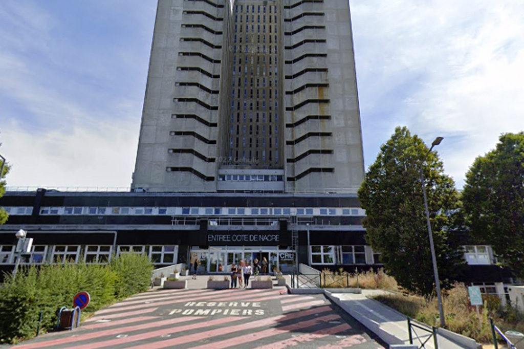 | |
Intensive care Mother-of-Pearl Coast Tower | Level : 20 | GPS access: Main | Parking: Parking 1 |  | |
Outpatient hospitalization Mother-of-Pearl Coast Tower | Level : 20 | GPS access: Main | Parking: Parking 1 | | |
Conventional Hospitalization · Heart / Vessels Mother-of-Pearl Coast Tower | Level : 20 | GPS access: Main | Parking: Parking 1 | | |
Heart Failure Treatment Unit Mother-of-Pearl Coast Tower | Level : 19 | GPS access: Main | Parking: Parking 1 | | |
Home-based clinical monitoring and follow-up of transplant patients Mother-of-Pearl Coast Tower | Level : 19 | GPS access: Main | Parking: Parking 1 | |
