Thoracic surgery
Thoracic surgery
The thoracic surgery department treats patients with pulmonary, pleural, mediastinal, costal or diaphragmatic pathology requiring surgical intervention.
Consulting & Team
Thoracic Surgery
Dr. Boddaert: Tuesdays from 9:00 AM to 12:00 PM and Thursdays from 9:00 AM to 11:00 AM;
Dr. Le Rochais: Wednesdays from 9:00 AM to 11:00 AM
Information for making an appointment
02 31 06 53 26 and 02 31 06 44 53
Service composition
- 3 Hospital Practitioners
- 1 Junior Doctor
- 1 Internal
- 2 Medical Secretaries
- 16 Nurses
- 14 Caregivers
- 1 Health professional
- Laure Pasquer
- Hospital PractitionerThoracic surgery
- Hospital Practitioner – Associate Professor at Val-de-GrâceThoracic surgery
- Medical Secretary
- Medical Secretary
Our services
The Thoracic Surgery department at Caen Normandy University Hospital treats patients with:
- Pulmonary pathologies : Surgery for lung cancers, surgery for lung metastases, diagnostic lung biopsies and surgery for lung infections.
- Pleural pathologies : Exploration and treatment of pleural effusions, diagnostic and therapeutic surgery of pleural tumors, management of pneumothorax.
- Mediastinal pathologies : Diagnosis and surgery of mediastinal tumors: thymomas, schwannomas, cysts, teratomas, lymphomas and hematological malignancies, etc.
- Parietal pathologies : Surgery of the chest wall, ribs and diaphragm.
- Sympathectomies for Hyperhidrosis and Eurotophobia
- Chest trauma : rib fractures, flail chest, sternum fracture, diaphragmatic ruptures, traumatic hemo-pneumothorax, penetrating wounds, ballistic trauma.
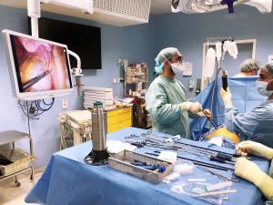
The department performs interventions via conventional approaches (thoracotomy, sternotomy) and also minimally invasive approaches (thoracoscopy and robot-assisted surgery), for pulmonary, pleural or mediastinal pathologies.
The practitioners work closely with the region's pulmonology departments and participate in the Multidisciplinary Thoracic Oncology Tumor Boards of the Caen Normandy University Hospital and the François Baclesse Cancer Center . They also regularly participate in national multidisciplinary tumor boards (MTBs ) on the management of thymic tumors (MTBs RYTHMIC) , as well as regional referral MTBs (MTBs , MTBs ). Sarcoma Endometriosis
The three practitioners in the department perform nearly 500 procedures per year, including more than 200 major lung resections (segmentectomies, lobectomies, bilobectomies or pneumonectomies).
- from a surgical consultation (unit 19-40),
- of an anesthesiologist consultation (level 1 consultation platform)
- of a hospitalization unit (unit 19-10),
- from an operating room,
- and a resuscitation-intensive care unit (unit 6-30).
Patient information sheets
PATIENT INFORMATION SHEET
– PNEUMOTHORAX –
What is a pneumothorax?
This is the presence of air in the thorax, between the lung and the inside of the ribs, in the pleural cavity. This air "detaches" the lung from the inside of the ribs.
What are the circumstances surrounding the occurrence of a pneumothorax?
Pneumothorax most often occurs spontaneously, at rest. In this situation, it is often due to small bubbles on the surface of the lung that rupture, air escaping from the lung and creating the pneumothorax.
Other contributing factors include trauma (falls, car accidents, etc.) or direct injuries (stab wounds, etc.). Exertion with a closed glottis (weightlifting, carrying heavy loads, playing wind instruments), by increasing intrathoracic pressure, can also contribute to the development of pneumothorax.
What are the risk factors for pneumothorax?
Spontaneous ( or primary/idiopathic ) pneumothorax typically occurs in young adults (15 to 35 years old). Smoking is the main risk factor for this condition. Regular cannabis use can also increase the risk of pneumothorax.
Being male and having a long, slender body type (tall and thin people) are also known risk factors.
In older patients, certain lung diseases such as emphysema (often caused by smoking) or chronic bronchitis can be responsible for pneumothorax.
Endometriosis can rarely cause pneumothorax in women. Generally, the pneumothorax is on the right side and occurs between the day before and 72 hours after the onset of menstruation. Specific management, most often involving surgery and a gynecological evaluation, is frequently necessary.
How is the diagnosis made?
Symptoms vary: chest pain, shortness of breath, a dry cough, and a feeling of breathlessness. Auscultation and questioning help guide the doctor. A simple chest X-ray is usually sufficient for diagnosis. A CT scan may be useful in certain circumstances.
What is the treatment for pneumothorax?
When the pneumothorax is well tolerated and the lung is only slightly detached, simple rest is often enough to bring the lung back into normal position.
When a pneumothorax is poorly tolerated and the lung is severely collapsed, the air trapped between the lung and the chest wall must be aspirated. This procedure is called " chest drainage ." It involves placing a tube (" drain ") between two ribs under local anesthesia.
If the pneumothorax resolves quickly (<5 days), the drain is removed. Otherwise, surgery is recommended.
What is the risk of recurrence?
The risk of recurrence after spontaneous pneumothorax is approximately 30 to 50%. This recurrence can occur in the weeks following or, conversely, several years after the first episode, without any possibility of prediction. Recurrence can occur on the same side, but also on the other side, as there are often blebs in both lungs.
This risk can be largely reduced by stopping cannabis and tobacco use.
When is surgery performed for a pneumothorax?
Surgical intervention is recommended in cases of recurrent pneumothorax or persistent air leak lasting more than 4 days
How is the intervention performed?
This procedure is performed using video-assisted thoracoscopy: three small incisions of approximately 2 cm are made on the side of the chest, allowing the operation to be performed with a camera. In rare cases, a larger incision may be necessary to access the area between two ribs.
The procedure involves removing a small portion of lung containing the blisters and creating adhesions between the inside of the ribs and the surface of the lung (symphysis). These adhesions can be achieved either by scraping the inside of the ribs (" pleural freshening ") or by introducing talc (" talc pleurodesis ").
A tube (" drain ") is inserted into the chest through one of the openings to collect secretions, bleeding, or air leaks after the operation.
What are the risks of surgery?
Air leaks may persist after the procedure, requiring the chest drain to remain in place for a few days. Bleeding may occur, but it is usually minor. Post-operative pain is managed with strong painkillers for a few days.
There is also a risk of pneumothorax recurrence, even after surgery, but this is low, less than 1%. These recurrences are most often partial and without consequences.
What precautions should be taken after a pneumothorax?
Physical activities, carrying heavy loads, and air travel are contraindicated during the first 3 weeks after a pneumothorax, whether or not it has been operated on.
Scuba diving with tanks is formally contraindicated for life, whether or not there has been intervention.
Beyond this 3-week period, no other activity is contraindicated.
Quitting smoking and cannabis is recommended at any age to prevent recurrences of pneumothorax.
Tobacco
Tobacco is responsible for numerous health problems:
| The University Hospital's Tobacco Cessation Unit, located on Level 1, can help you with your efforts to quit smoking or cannabis: 02 31 06 49 82 Ask to meet with the tobacco cessation specialists during your hospitalization! |
- Arteritis
- Heart attack
- Strokes
- Lung cancers
- Bladder cancers
- Pneumothorax
- Pulmonary emphysema
- Chronic bronchitis
… (non-exhaustive list!)
PATIENT INFORMATION SHEET
– PLEURAL BIOPSIES AND TALCAP –
What is the purpose of the intervention?
There are several advantages to performing pleural biopsies and talc pleurodesis
- Symptomatic: Fluid around the lung compresses it and can cause several symptoms: difficulty breathing, dry cough, abnormal sensations in the chest. Draining the fluid relieves these symptoms.
- Diagnosis: The cause of fluid production is not always determined before surgery. Performing biopsies (sampling) usually reveals why fluid has accumulated around your lung. If the cause is known, further biopsies may allow for additional analysis and, in some cases, modify the treatment of the underlying condition.
- Treatment: Applying talc can prevent the re-accumulation of fluid after surgery. This eliminates the need for pleural taps, and symptoms related to fluid around your lungs will disappear or be alleviated.
How is the intervention performed?
This procedure requires general anesthesia
This procedure is generally performed using video-assisted thoracoscopy: two or three small incisions of about 2 cm are made in the side of the chest, allowing the operation to be performed with a camera. Rarely, a larger incision may be necessary to access the area between two ribs.
- The procedure involves removing the fluid present in the pleural cavity (space between the ribs and the lung) when there is any.
- Next, the entire thoracic cavity can be inspected using the camera and pleural biopsies (= of the pleura), generally on areas with abnormal appearance.
- Talc is introduced at the end of the procedure. Its purpose is to create adhesions between the inside of the ribs and the surface of the lung (symphysis), to prevent the re-production of fluid.
A tube (" drain ") is inserted into the chest through one of the openings to collect secretions, bleeding, or air leaks after the operation.
How long does the surgery take?
This type of procedure lasts approximately 20 to 30 minutes
How long does one stay in the hospital?
The patient returns the day before or the morning of their procedure.
At the end of the operation, a drain is left in place, usually for a period of one to two days, but sometimes a little longer.
You can go home the day after the drain is removed, after a follow-up X-ray.
The length of hospital stay is therefore generally 4 to 5 days.
What are the risks of surgery?
Air leaks or significant fluid production may occur after the procedure, requiring the chest drain to remain in place for a few days. Bleeding may also occur; it is usually minor. Post-operative pain is managed with strong analgesics for a few days.
There is often an area on the front of the chest, under the breast, that is slightly numb for a few weeks due to the surgical incisions.
Is the intervention still working?
In most situations, pleural biopsies allow for diagnosis and understanding of the origin of fluid production.
However, the effectiveness of talc pleurodesis is less certain : For talc to "work" and prevent fluid from regenerating, the lung must reinflate. When there is fluid around the lung, it deflates, and sometimes a capsule forms on its surface. During the operation, an attempt is made to reinflate the lung, but the capsule sometimes prevents it from regaining its normal volume. It is impossible to know whether the lung will reinflate properly before the operation.
- If the lung does not reinflate properly, talc pleurodesis may only be partially effective. In these situations, further pleural taps may be necessary.
- If the lung reinflates properly, then talc pleurodesis has a high chance of working.
Your surgeon, as well as the nursing team of the department, are available to answer your questions and supplement this general information, which does not prejudge your specific care, related to your pathology or your medical history.
The ERAC booklet for the department is given to all patients scheduled for surgery in the department.
It provides a lot of information about the course of the stay, but also about the post-operative period and the possible risks and complications related to the different surgeries.
This booklet provides supplementary information for patients and their families, in addition to the information given by practitioners during consultations
Structure | Center page |
Locate / contact the service
Côte de Nacre Hospital
Thoracic surgery
Secretariat of the department
| Level | GPS access | Entrance | |||
|---|---|---|---|---|---|
Thoracic surgery Côte de Nacre Hospital | Level : 19 | GPS access: Main | |